Ultra-Fast holographic microscope for viewing the nerve network
Acquires optical information 100 times faster and allows the observation of the living nerve network without fluorescent labels

▲ Professor Wonshik Choi of the Department of Physics at the College of Science
A research team led by Wonshik Choi (professor of Korea University), the associate director of the Center for Molecular Spectroscopy and Dynamics, Institute for Basic Science (IBS, President Doochul Kim), developed an ultra-fast holographic microscope that allows the observation of the nerve network of living organisms at a high resolution without a surgical operation.
Inner biological tissues are complicated structures that are difficult to observe, even with a microscope, because the light wavefront is distorted as the light collides with various types of cells. A holographic microscope was developed in order to overcome this limitation of general optical microscopes for in-depth observation of biological tissues. Acquisition of deep-depth biological images requires selectively measuring specific depths by quantifying the light wavefront. In contrast to general microscopes that observe only the light intensity, a time-resolved holographic microscope simultaneously measures the intensity and the phase of light by using two types of light (laser), which are a reference light and an object light. Based on the measured light intensity and phase, optical signals are selectively acquired at a specific depth to obtain the images from deep in the tissue.
* Wavefront: The surface obtained by connecting all points where the wave has the same phase. For example, the wavefront of a wave formed by throwing a stone into a calm lake is circular.
* Time-resolved holographic microscope: Holographic microscopy is a technology for measuring the amplitude and phase of light based on the optical interference between two laser lights. In particular, a time-resolved holographic microscope allows one to selectively acquire optical signals at a specific depth by using light sources causing a very short interference of about 10 ㎛.
However, conventional technology requires repeated hardware-based wavefront measurement and control in order to overcome the wavefront distortion, and thus is difficult to apply to the observation of living animals due to the slow image acquisition speed.
Professor Choi’s research team improved the data acquisition speed by an order of magnitude in comparison with the conventional technology by synchronizing the object light with the reference light. While the image acquisition speed of the conventional technology is about 10 images per second, that of the ultra-fast holographic microscopy developed by his team is about 500 images per second.
The technology also allowed an increase in the amplitude of the optical signals at the focus, even without the repeated hardware-based processing to measure and control the wavefront. This means that the performance for correcting the wavefront distortion has been improved by more than 100 times for in-depth observation.
The research team used the ultra-fast holographic microscope to successfully acquire high-resolution cranial nerve network images from the posterior brain of a living zebrafish without using a fluorescent label. Most existing optical microscopy technologies require the injection of a fluorescent substance into a young zebrafish within one week after hatching to investigate the structure of nerve fibers. As a zebrafish grows, the scales that cover the posterior brain make it difficult to view inside the brain. On the contrary, the newly developed technology allows the acquiring of label-free high-resolution images of the nerve network of the central nervous system from a zebrafish that is several weeks old.
Professor Wonshik Choi of the Department of Physics, associate director of the Center for Molecular Spectroscopy and Dynamics, said, “Our technology has overcome the depth limitation of the conventional optical microscopy technologies.” He added, “This technology will make a great contribution to not only the brain and nerve sciences but also various bio-convergence studies and industries requiring precise measurement.”
The results of the study were published online in the international journal, Nature Communications (IF 11.878), July 17 issue.
* Title of Article: “Label-free neuroimaging in vivo with adaptive opticalsynchronous angular scanning microscopy”/Nature Communications
* Authors: Moonseok Kim, Yonghyeon Jo, Jin Hee Hong, Suhyun Kim, Seokchan Yoon, Kyung-Deok Song, Sungsam Kang, Byunghak Lee, Guang Hoon Kim, Hae-Chul Park and Wonshik Choi
[Description of Figures]
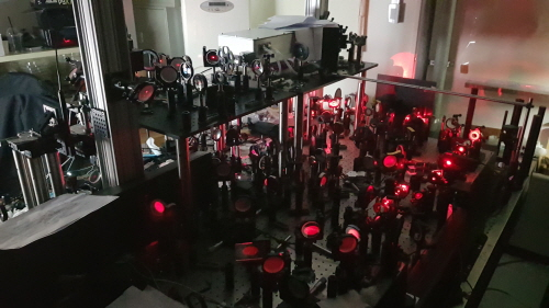
▲ Figure 1: An ultra-fast holographic microscope.
An ultra-fast holographic microscope developed by a research team from the Center for Molecular Spectroscopy and Dynamics, Institute for Basic Science. The newly developed microscope has an image acquisition speed higher by an order of magnitude than that of conventional microscopes, and thus allows the in-depth observation of the nerve networks of living organisms.
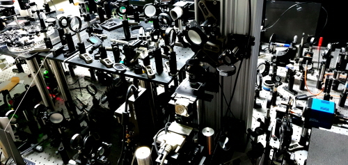
▲ Figure 2: An ultra-fast holographic microscope.
An ultra-fast holographic microscope developed by a research team from the Center for Molecular Spectroscopy and Dynamics, Institute for Basic Science. The newly developed microscope has an image acquisition speed higher by an order of magnitude than that of conventional microscopes, and thus allows the in-depth observation of the nerve networks of living organisms.

▲ Figure 3: Principle of a time-resolved ultra-fast holographic microscope.
The time-resolved ultra-fast holographic microscope (a) uses a scanning mirror to simultaneously alternate the light that illuminates an object and the reference light for recording the returning interference pattern. While the conventional technology for scanning the object light with a fixed reference light allows observation only within a limited area, the newly developed technology (C) can be used to record the interference pattern in all areas.
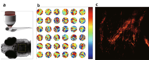
▲ Figure 4: Technology for correcting wavefront distortion in living tissues.
In a zebrafish 21 days after hatching, it is difficult to observe the inside of the posterior brain by using the conventional technology because the brain area is covered with thick scales (a). The research team was able to numerically detect the wavefront distortion of the light in each area (b) and clearly observe the disentangled fibrous structure of the nerve system.
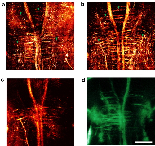
▲ Figure 5: Three-dimensional observation of the nerve network in the posterior brain of a living zebrafish.
The change in the nerve network structure in the central nervous system depending on the development stage was observed in a zebrafish six days (a) and ten days (b) after hatching. The performance comparison of the microscopes revealed that the reflectance image (c) and the fluorescent image (d) of a zebrafish 10 days after hatching, obtained by using a general confocal microscope, showed the nerve system but not the high-resolution nerve fiber structure.